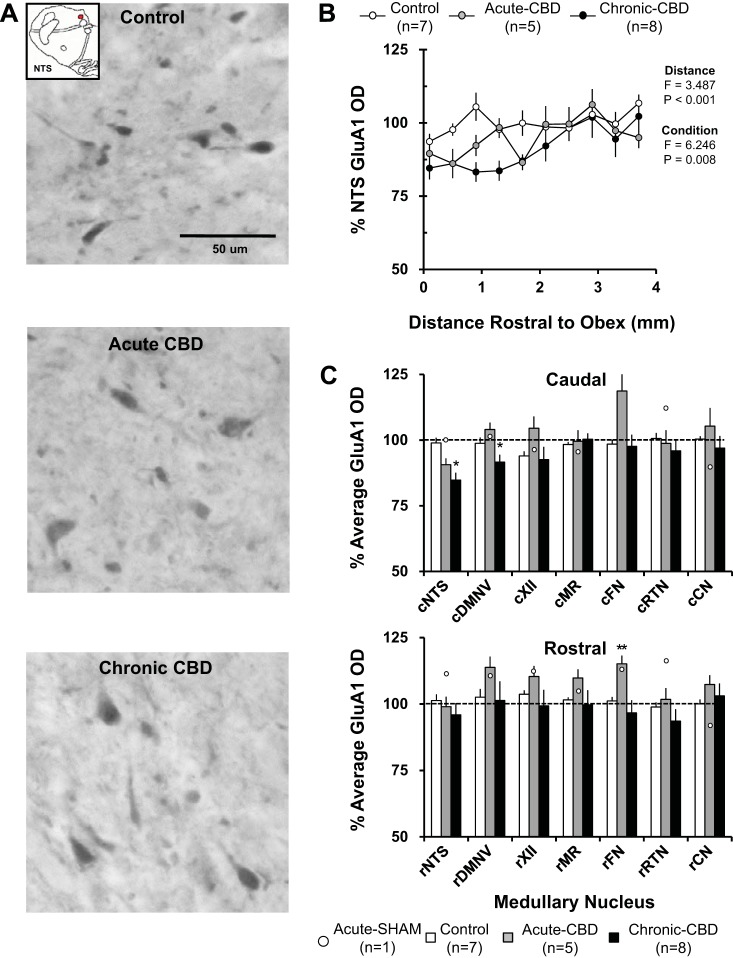
Fig. 5.
GluA1 immunoreactivity demonstrated site-specific effects both acutely and chronically after CBD. A: GluA1-ir neurons within the NTS (20X, 0.50 NA) of a naive goat (top panel), a goat euthanized during hypoventilation after CBD (acute CBD-5 days; middle panel), and a goat euthanized after partial recovery of resting PaCO2 (chronic CBD-30 days; bottom panel). Note there were minimal qualitative decreases in GluA1 immunoreactivity in the acute CBD goat and chronic CBD goat. B: GluA1 optical density quantification within the NTS. On the y-axis is plotted normalized GluA1 OD ± SE, and on the x-axis is plotted distance rostral from obex (mm) in naive/sham-CBD goats, acute-CBD goats, and chronic CBD goats. The F and P values are from the 2-way ANOVA (distance and condition as factors) on these data. C: average normalized GluA1 OD ± SE in 6 different respiratory nuclei and one nonrespiratory nucleus, the cuneate nucleus, in naive/sham-CBD goats, acute CBD goats, and chronic-CBD goats. C, top panel, shows GluA1 OD from the caudal (c) division of each nucleus, and C, bottom panel, shows the rostral (r) division. The open symbols represent GluA1 OD from the individual sham-CBD goat euthanized at 5 days post-sham surgery. *P < 0.05 vs. naive/sham-CBD, **P < 0.05 vs. naive/sham-CBD and chronic CBD. DMNV, dorsal motor nucleus of the vagus nerve; XII, hypoglossal motor nucleus; MR, medullary raphé; FN, facial motor nucleus; RTN, retrotrapezoid nucleus (region ventral to the FN); CN, cuneate nucleus.

