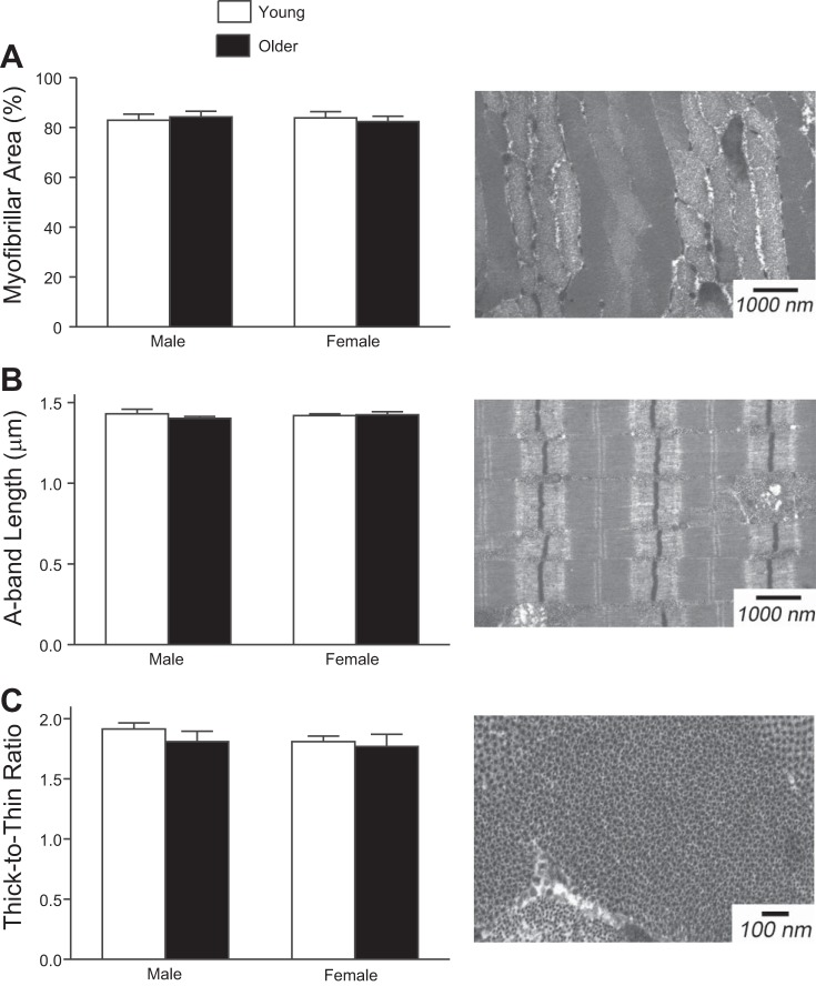
Fig. 3.
Myofibrillar fractional area (A), A-band length (B), and thick-to-thin-filament ratio (C) were assessed using transmission electron microscopy (EM). These indexes of muscle ultrastructure were not different between young and older men and women. Example images from a young woman are presented to the right of the corresponding data.

