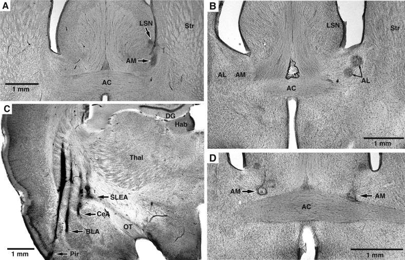
Fig. 1.
Histological identification of recording sites. A–D: coronal sections counterstained with cresyl violet. Recording sites are marked with small electrolytic lesions (arrows). A: electrolytic lesions in the lateral septal nucleus (LSN) and bed nucleus of the stria terminalis (BNST) medial site (AM). B: 2 recording sites in BNST lateral site (AL). C: recording sites in the piriform (Pir) cortex, basolateral complex of the amygdala (BLA), central amygdala (CeA), and sublenticular extended amygdala (SLEA). Too few BLA and Pir recordings were obtained for meaningful analyses. Data obtained in CeA and SLEA were pooled since they are thought to constitute an anatomic entity. D: 2 recording sites in the left and right BNST-AM. AC, anterior commissure; DG, dentate gyrus; Hab, habenula; OT, optic tract; Str, striatum; Thal, thalamus.

