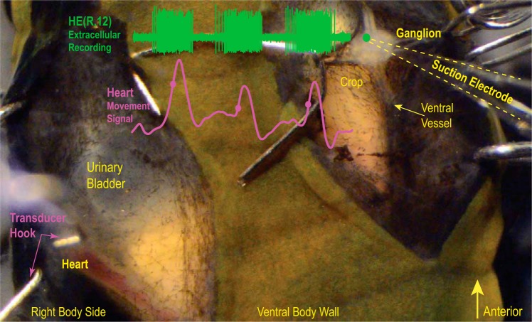
Fig. 2.
Minimally dissected leech. Ventral view of segment 12. Small hooks on both sides held the preparation in place. The exposed ganglion is seen on the right, with the green dot showing the approximate position of the HE heart motor neuron in the ganglion. The bursting activity of the heart (HE) motor neuron was recorded extracellularly (green trace), and heart movements were recorded with a movement transducer (pink trace). Filled circles indicate the MRR of the movement signal. The hook of the movement transducer cradles the heart. Note that the heart is filled with blood.

