Fig. 6.
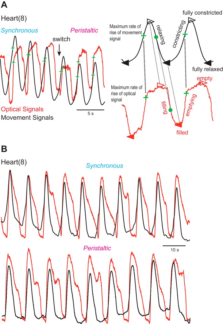
Alignment of optical signals (red traces) and movement signals (black traces) in minimally dissected animals. Optical and movement signals were vertically matched in size to facilitate comparison. A: recordings are across a switch from the synchronous to the peristaltic mode in heart(8). The 2 expanded beat cycles on right show the 4 time points that the analysis code identified for the movement and the optical signals, respectively: filled triangles correspond to the trough (i.e., a relaxed and filled heart), open triangles to the peak (i.e., a constricted and empty heart), green crosses to the MRR (i.e., constricting and emptying), and filled circles to the maximum rate of decay (i.e., relaxing and filling). Note that constricting (upstroke of the movement signal) and emptying (upstroke of the optical signal) align well and that the MRR of the movement signal corresponds to the MRR of the optical signal. B: same experimental design, different animal, heart(8). Note that optical and movement traces align perfectly during constricting and emptying in both coordination modes but that filling is delayed and the heart remains empty for some time while relaxing.

