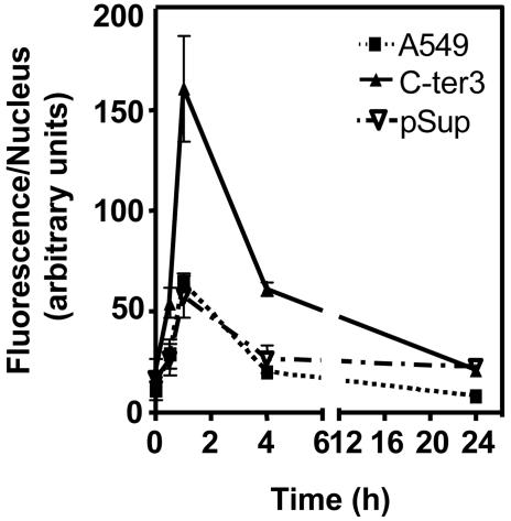
Fig. 3.
Quantification of the average integrated fluorescence intensity per nucleus due to phosphorylation of histone H2AX as a function of time after 5-Gy irradiation. [Fig. 6 shows cells stained with a nuclear stain (4′,6-diamidino-2-phenylindole) and Abs to phosphorylated histone H2AX (γ-H2AX) at various times after irradiation.]

