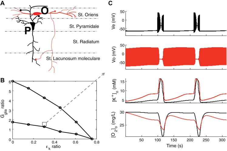
Fig. 7.
Interplay between pyramidal cell and inhibitory interneuron. A: structure of the hippocampal CA1 2-neuron model. The cell body of pyramidal cell (P, black) in stratum (st.) pyramidal layer and the cell body of oriens lacunosum moleculare (OLM; O, red) interneuron in the st. oriens layer. B: region for the alternating interplay in firing between 2-neuron model. When the ratio of Gglia and oriens OLM, calculated as OLM/pyramidal, are in the region bounded by the 2 lines, the 2 cells exhibit firing interplay. The ratios are calculated when the parameters of the pyramidal cell are fixed as in Table 1. An example of the interplay in the region of the square is shown in C. In C, the dual-layer model shows a nearly identical electrical and [O2]o interplay as seen experimentally (Žiburkus et al. 2006, 2013, Ingram et al. 2014, companion article). When the OLM cell (Vo, red) goes into depolarization block the pyramidal cell (Ve, black) is released and exhibits runaway excitation behavior until the OLM cell resumes firing. Extracellular potassium concentrations of pyramidal cell (black) and OLM cell (red) are elevated during seizure events. Even though the OLM cell fires more than the pyramidal cell, local oxygen concentration around cell body in st. pyramidale (black) is lower than in st. oriens layer (red), which is due to the higher packing density of pyramidal cells. Note, importantly, that the maximal O2 consumption occurs during depolarization block in the model, consistent with experimental observations, reflecting maximal stimulation of Na-K pump under conditions of maximal gradient collapse.

