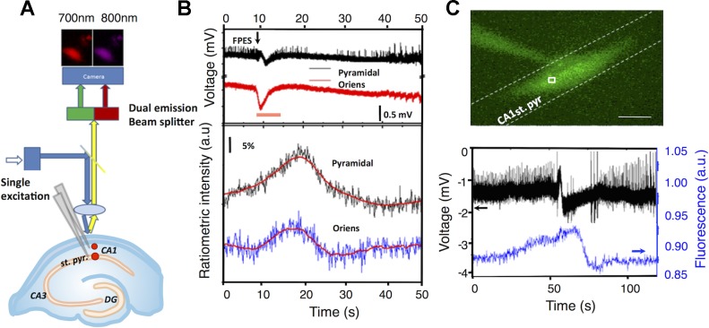
Fig. 1.
Probes encapsulated by biologically localized embedding (PEBBLE) and liposome nanosensing during spontaneous seizure events. A: PEBBLE nanosensors were injected into the CA1 stratum (st.) pyramidale and st. oriens. Absorption at 450 ± 30 nm was used to excite the reference (HiLyte 680) and oxygen sensitive dye (Oxyphor G2) and the emission beam was split into two wavelengths (700 ± 30 and 800 ± 40 nm) through a dual emission beam splitter and captured on a single CCD camera. Simultaneous electrical activity was recorded using local field potential electrodes in the st. oriens and st. pyramidale. B: ratiometric intensity (bottom) and local field potential recordings (top) during a 4-aminopyridine (4-AP)-induced spontaneous seizure-like event (orange bar) in the CA1 st. oriens and st. pyramidale (n = 2). C: picoinjection of liposomes within CA1 st. pyramidale. Optical recordings were monitored over a 25 × 25 μm region of interest. Scale bar = 100 μm. Simultaneous recording of neuronal activity and Ru(phen)3 intensity within the CA1 pyramidal layer during 4-AP-induced spontaneous seizure event, demonstrating a decrease in oxygen in advance of the electrical correlate of seizure-like event (n = 3). FPES, fast positive extracellular shift; DG, dentate gyrus; a.u., arbitrary units.

