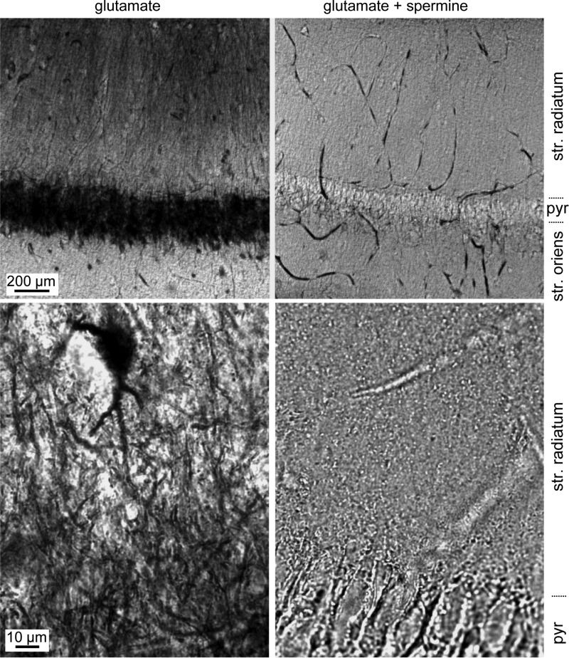
Fig. 3.
Loading of CA1 cell dendrites with Co2+. Acute hippocampal slices were submerged in saline containing 5 mM CoCl2 and 10 mM glutamate for 20 s to activate AMPARs, followed by 40 s in saline containing Co2+ alone, repeated 10 times over 10 min. Slices were then fixed, resectioned, and stained for the presence of Co2+ using silver intensification. Low-magnification images of area CA1 (top row) and high-magnification images of s. radiatum (bottom row) of slices treated with glutamate alone (left column) or glutamate and spermine (right column). Intense labeling was seen in the somata and dendrites of slices exposed to glutamate, and this staining was absent when glutamate was applied in the presence of spermine. A heavily stained s. radiatum interneuron is visible in the high-power image at left. pyr, S. pyramidale.

