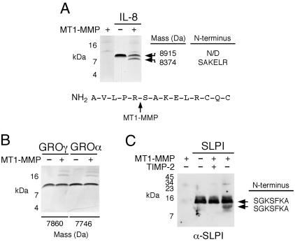
Fig. 2.
MT1-MMP processing of IL-8 and SLPI. (A–C) CXC chemokines or SLPI were incubated with sMT1-MMP for 18 h at 37°C. Samples were then separated on 15% Tris-tricine gels under nonreducing conditions. (A) MT1-MMP cleavage of IL-8 and sequence of the cleavage site. (B) MT1-MMP did not cleave GRO-α or -γ as analyzed by SDS/PAGE. MALDI-TOF MS revealed identical masses for both chemokines in the presence or absence of MT1-MMP. (C) SLPI was detected by Western blotting by using α-SLPI antibody. SLPI cleavage was blocked by TIMP-2 added with sMT1-MMP or after preincubation (not shown). N-terminal sequences are shown.

