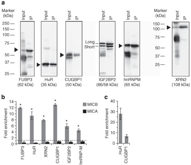
Figure 2. The mRNA of MICB endogenously associates with the identified RBPs.
RIP analysis was performed with RKO whole-cell lysate. (a) Whole membranes of WB analysis of the immunoprecipitation (IP) of the specific RBPs. Input is total protein input used for the RIP experiment, IP is the sample in which a specific antibody was used (b) Fold enrichment of the mRNAs of MICB (grey) or MICA (black) bound by the specific RBP (x axis) as assessed by qRT-PCR. Shown are mean ± s.e.m. of triplicates. Data are representative of three independent experiments performed. *P<0.003 by paired Student’s t-test of all three independent experiments combined. (c) qRT-PCR of known transcripts bound by HuR (β-actin) or CUGBP1 (c-JUN). Shown is mean fold enrichment of the known target relative to total RNA of two independent experiments ± s.d.

