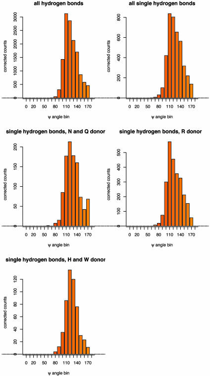
Fig. 4.
Distributions of the acceptor angle Ψ for hydrogen bonds observed in high-resolution protein crystal structures. Shown are the distributions for all side-chain–side-chain hydrogen bonds with sp2 hybridized acceptor atoms in the data set of 698 protein crystal structures, a subset of those hydrogen bonds where only a single hydrogen bond is made to each acceptor atom, and subsets of the single hydrogen bonds split by the type of the donor amino acid (R, arginine; N, asparagine; Q, glutamine; H, histidine; W, tryptophan). Raw counts were corrected for the different volume elements encompassed by the bins; the angular correction is sin(Ψ).

