Fig. 1.
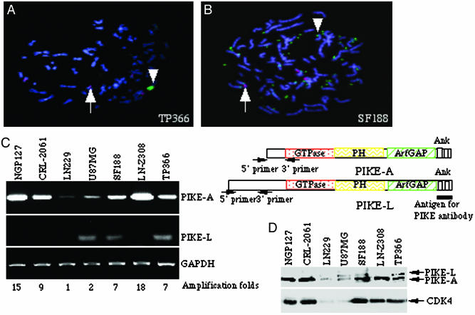
PIKE-A is amplified in human glioblastoma cells. (A and B) Fluorescent in situ hybridization staining of glioblastoma TP366 (A) and SF188 (B) cells. Red dot stains for the centromere of human chromosome 12, whereas the green stains for PIKE-A with the amplified PIKE (arrowhead) versus normal PIKE (arrow). Amplified PIKE-A translocates from chromosome 12 to another chromosome in TP366 cells (A). Amplified PIKE-A also forms double minute chromosomes in SF188 cells (B). (C) RT-PCR analysis of PIKE-A and -L mRNA expression in human cancer cell lines. PIKE-A mRNA is robustly expressed in NGP-127, CRL-2061, TP366, LN-Z308, and SF188 cells, but not in LN229 or U87MG cells (Top, left lane). By contrast, PIKE-L is weakly expressed in U87MG, SF188, and TP366 cells (left lane, Middle). Equal levels of GAPDH were monitored as control (Bottom, left lane). The relative gene amplification folds are labeled at the bottom. The primers for PIKE-A and -L used in RT-PCR analysis and the antigen used to raise anti-PIKE antibody are indicated on the right. (D) Western blotting analysis of PIKE-A protein expression. Cell lysate (50 μg) of a variety of human glioblastoma cell lines was used for immunoblotting analysis with rabbit polyclonal anti-PIKE and mouse monoclonal CDK4 antibodies.

