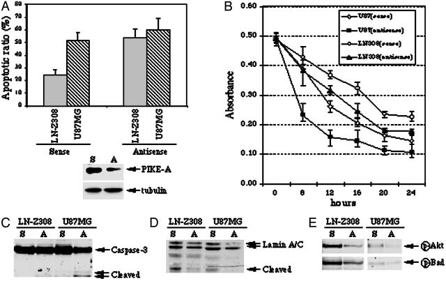
Fig. 5.
PIKE-A knockdown increase apoptosis by inactivating Akt. (A) Apoptotic assay. Serum-starved cells were treated with Penetratin 1-conjugated antisense and sense oligonucleotides of PIKE-A. After 6 h, the cells were treated with staurosporine for 16 h, and followed by chromatin condensation and fragmentation analysis with DAPI staining (Upper). PIKE-A expression is selectively decreased by antisense but not control sense oligonucleotide. By contrast, tubulin expression level is not changed (Lower). (B) MTT cell viability assay. Serum-starved cells were treated with Penetratin 1-conjugated antisense and sense oligonucleotides of PIKE-A. After 6 h, the cells were treated with staurosporine for various time points, and followed by MTT assay. (C and D) Western blotting analysis of Caspase-3 and Lamin A/C cleavage in sense and antisense oligonucleotide treated cells during apoptosis. (E) PIKE-A knockdown inhibits Akt activity. Western blotting analysis of Akt and Bad phosphorylation in sense and antisense oligonucleotide-treated cells. Consistent with PIKE-A knockdown, Akt phosphorylation is diminished in antisense but not sense oligonucleotide treated cells (Upper). The phosphorylation of Akt physiological substrate, Bad, in sense and antisense oligonucleotide-treated cells (Lower).

