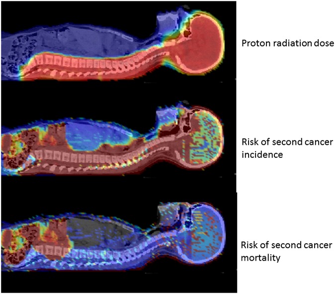
Figure 10.
Dose and risk distribution for second cancer. Images of a 9-year old girl who received craniospinal irradiation for medulloblastoma using passively scattered proton beams at MD Anderson Cancer Center. The colour scale illustrates the difference for absorbed dose, incidence and mortality cancer risk in different organs. Radiation absorbed dose depends strongly on patient anatomy and treatment factors. Risk of second cancer incidence and mortality varies strongly with radiation dose, but, importantly, it also varies strongly between organs, with the patient's age at exposure and attained age, sex, genetic profile, and other factors. Reproduced from Newhauser and Durante108 with permission from Nature Publishing Group.

