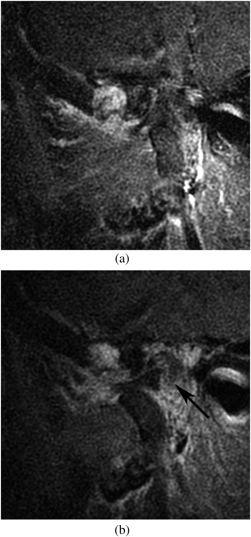
Figure 3.
MR images of the medial part of the temporomandibular joint. (a) The contrast-enhanced T1 weighted image shows no continuity between the lesion in the anterior of the condylar head and the eminence in the closed position. (b) Area in the posterior of the condylar head showing a low signal intensity on contrast-enhanced T1 weighted image (arrow).

