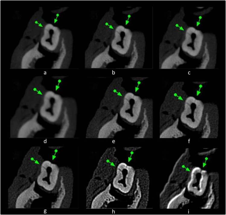
Figure 1.
Axial cone beam CT slices show the longitudinal root fracture (indicated by arrows) in first mandibular molar with (a) no filter, (b) Sharpen Mild, (c) Sharpen Super Mild, (d) S9, (e) Sharpen, (f) Sharpen 3 × 3, (g) Angio Sharpen Medium 5 × 5, (h) Angio Sharpen High 5 × 5 and (i) Shadow 3 × 3.

