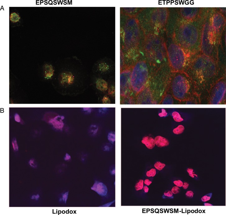
Fig. 3.
(A) Cellular localization of phage EPSQSWSM (left) and ETPPSWGG (right) in PANC-1 cells. PANC-1 cells were treated with phage, fixed, permeabilized, treated with anti-fd phage IgG, stained with an Alexa Fluor 488 conjugated goat anti-rabbit IgG and were visualized with a FITC filter under a confocal microscope. (B) Microscopy of doxorubicin uptake by PANC-1 cells delivered by unmodified Lipodox (left) compared with EPSQSWSM-Lipodox (right).

