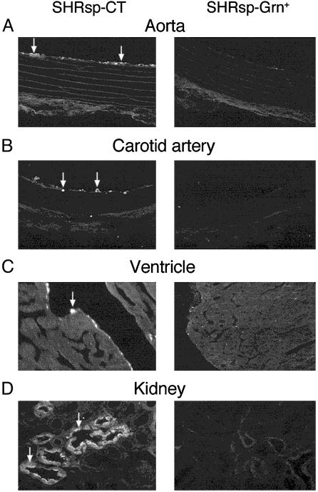
Fig. 3.
Representative micrographs demonstrating localization of activated macrophages (arrows indicate bright fluorescent cells) in the inner tissue linings of the aorta (A), carotid artery (B), and ventricle of the heart (C), as well as in the medulla of the kidney (D) in 19 week-old SHRsp on CT or Grn+ diet. Few such activated macrophages are present in tissues of animals on Grn+ diet.

