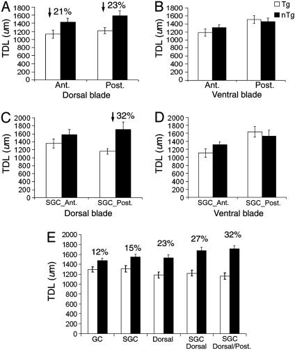
Fig. 4.
Comparison of the TDL of GCs across the A-P axis between Tg and nTg mice. (A) Consistent reductions in TDLs of GCs within the dorsal blade by A-P axis. A 21% reduction was found anteriorly, whereas a 23% reduction was found posteriorly. Ant., anterior; Post., posterior. (B) No significant differences were found in the ventral blade across the A-P axis. (C) A prominent reduction occurred exclusively in SGCs of the dorsal blade across the A-P axis (DGC data not shown). The analysis has further localized a significant reduction (32%) in TDLs only in the posterior pole. (D) No significant differences were found in SGCs of the ventral blade along the A-P axis. (E) Localization of selective dendritic pathology of GCs in 90-d Tg mice.

