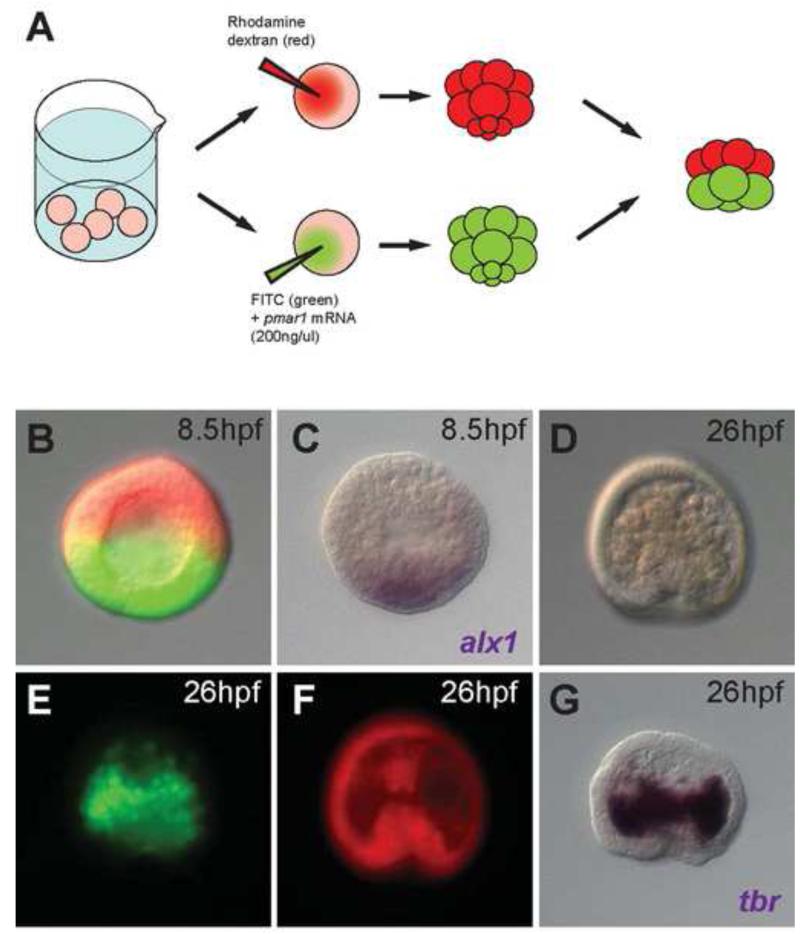
Figure 2.
(A) Experimental procedure for obtaining micromere(−) embryos overexpressing pmar1 in macromeres. (B) A micromere(−) embryo overexpressing pmar1 in its macromeres, with the animal cap cells labeled by rhodamine dextran (red) and progeny of pmar1-injected macromeres labeled by FITC (green). (C) alx1 in situ hybridization for the same embryo in (B) indicating the activation of alx1 in half of the embryo. (D-G) A sibling embryo of (B) developed to 26hpf. (D) The gastrulated embryo showed increased number of ingressed cells in the blastocoel. (E) The ingressed cells derived almost entirely from pmar1-injected macromere labeled with FITC (green) and started to aggregate on the sides of the archenteron. (F) Animal cap cells labeled with rhodamine dextran (red) contributed to the archenteron. (G) In situ hybridization of tbr for the same embryo in (D-F) showing that the FITC-labeled cells (green) were tbr expressing skeletogenic cells.

