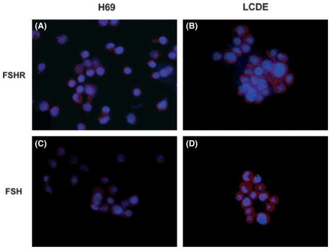
Fig. 5.
Representative immunofluorescence for (A, B) FSH receptor FSHR and (C, D) follicle-stimulating hormone FSH in H69 and LCDE human cell lines. In the LCDE cells, the expression of FSHR and FSH is higher when compared with the non-malignant cholangiocytes (red), whereas in blue are stained nuclei (DAPI). No staining was visible when primary antibodies were replaced with non-immune serum. Bar = 200 μm.

