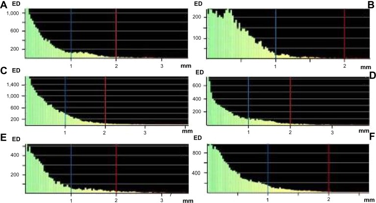
Figure 7.
Surface-deviation analysis for the shape of the mandible reconstruction with the iliac crest bone flap and osteomyocutaneous fibula flap.
Notes: Iiliac crest bone flap: Iliac flap shape (A), right mandible shape (B) and left mandible shape (C). Osteomyocutaneous fibula flap: fibula flap shape (D), right mandible shape (E) and left mandible shape (F). The calculation showed a surface deviation <1 mm (blue line) and <2 mm (red line).
Abbreviation: ED, element distribution.

