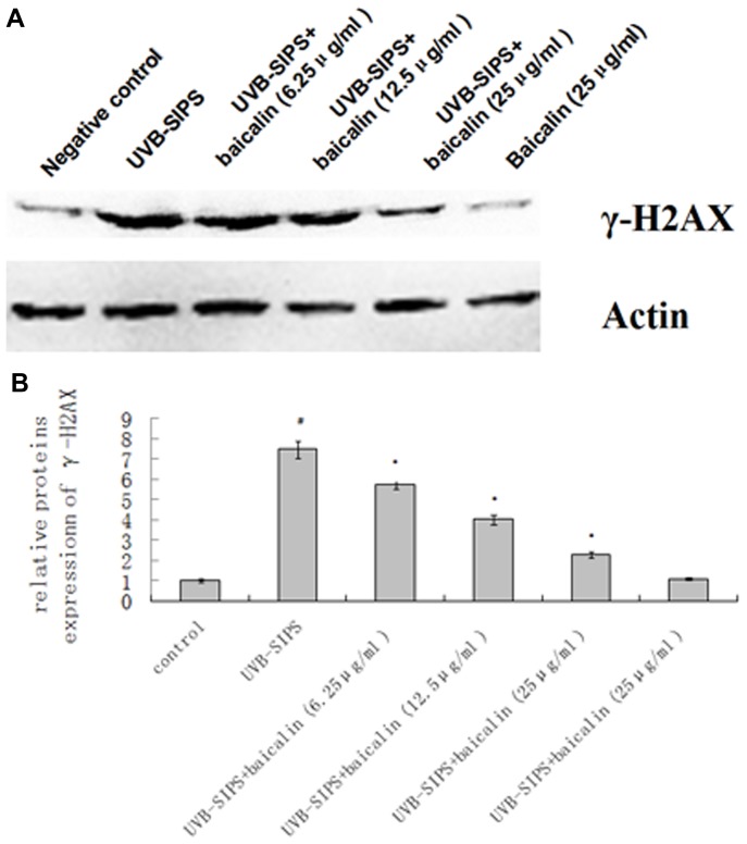
Figure 11. Baicalin protects cultured HDFs against UVB-SIPS induced expression of γ-H2AX proteins.
(A) Expression of γ-H2AX after UVB stress treated or untreated with baicalin were detected by Western blotting. (B) Band-Scan software was used to analyze each band in the gray scale. The ratio of gray scale values represents the ratio of amount of protein of interest/actin, and we calculated the relative ratio of every treatment group/control group. The results represented the mean relative ratio of triplicates. Statistical analysis was carried out with Student's t-test. The symbol (#) indicates a significant difference (p<0.05) between the control group and the UVB-SIPS group. Asterisks (*) indicate significant differences of p<0.05, respectively, between the baicalin-treated and UVB-SIPS cells.

