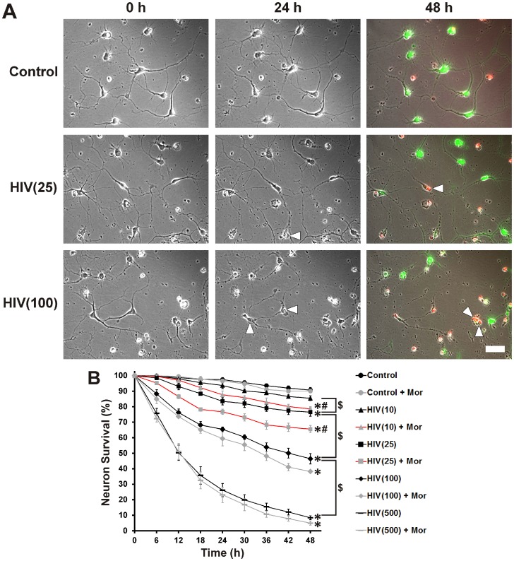
Figure 2. Concentration-dependent neuronal death in cultures treated with HIV+ sup ± morphine.
Individual striatal neurons were selected prior to treatment and repeatedly imaged for 48(A) Digital images of the same cells/fields, at 0 h, 24 h and 48 h after treatment (white arrowheads indicate cells that have died over the previous 24 h period). Live and dead cells were confirmed at the end of the experiment by staining respectively with calcein-AM (green) and ethidium homodimer-1 (red); scale bar = 40 µm. (B) Cells were assessed for viability at 6 h intervals in digital images. Findings were reported as the average neuronal survival as a percent of pre-treatment neuron count ± SEM. Significance was analyzed by repeated measures ANOVA and Duncan's post hoc test, from n = 3 separate experiments (at least 150 neurons per treatment group). Over the period of 48 h, all treatment groups except morphine alone [Control + Mor] and p24 = 10 pg/ml of HIV+ sup alone [HIV(10)], showed significantly reduced neuronal survival (*p<0.05 vs. Control). Neuronal survival declined in a concentration dependent manner with HIV+ sup treatment ($ p<0.05). Morphine showed significant interaction with HIV+ sup, but only at lower levels of exposure (p24 = 10 and 25 pg/ml) (# p<0.05 vs. HIV+ sup alone at corresponding titer). Control = Controlsup; HIV = HIV+ sup (concentration of p24 in pg/ml is specified in parentheses); Mor = morphine sulfate (500 nM).

