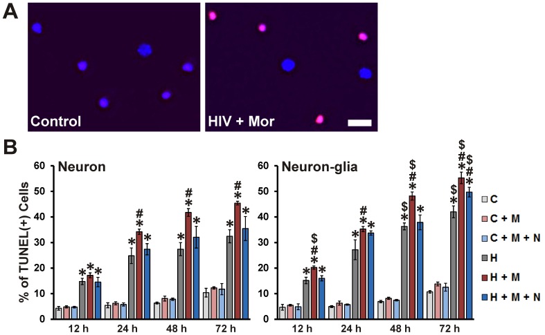
Figure 3. Neuronal apoptosis induced by HIV+ sup ± morphine.
Cells were fixed at specific intervals after treatment and labeled for Hoechst 33342 (blue) and TUNEL (red). (A) Digital images of neuronal cultures at 72 h after treatment; scale bar = 40 µm. (B) Apoptosis was assessed by manually counting the percentage of TUNEL(+) cells. Findings were reported as the average percentage of TUNEL(+) cells ± SEM. Significance was analyzed by one-way ANOVA and Duncan's post hoc test, from n = 4 separate experiments. At all assessed time points, in both culture systems, all groups exposed to HIV+ sup showed significantly enhanced neuronal apoptosis (*p<0.05 vs. respective C group). In all cases, except at 12 h in cultures with neurons alone, morphine significantly augmented HIV+ sup-mediated neuronal apoptosis (# p<0.05 vs. respective H group). In all cases, except for 72 h in neuron-glia cultures, the interactive effects of morphine were significantly attenuated by naloxone. In most cases, the presence of glia significantly enhanced HIV+ sup ± morphine-mediated neuron apoptosis ($ p<0.05 vs. corresponding treatment in neuron cultures; compare panels). C = Controlsup; H = HIV+ sup (p24 = 25 pg/ml); M = morphine sulfate (500 nM); N = naloxone (1.5 µM).

