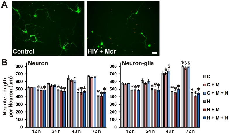
Figure 5. HIV+ sup ± morphine-mediated neurite damage.
Cells were fixed at specific intervals after treatment and labeled for MAP-2 (green) and TUNEL (red). (A) Digital images of neuronal cultures at 72 h after treatment; scale bar = 40 µm. (B) The ‘Sholl score’ was assessed only for TUNEL(-) neurons in the digital images and converted into neurite length in µm via a micrometer-scale calibration. The findings were reported as average total neurite length per neuron (µm) ± SEM. Significance was analyzed by one-way ANOVA and Duncan's post hoc test from n = 4 separate experiments. At all time-points and in both culture systems, all groups exposed to HIV+ sup showed significantly reduced neurite length (*p<0.05 vs. C). Morphine did not show a significant interaction with HIV+ sup treatment. The presence of glia did not have a significant effect on HIV+ sup ± morphine-mediated neurite damage, but in the presence of glia, Controlsup-treated groups showed significantly longer neurite length ($ p<0.05 vs. corresponding treatment in neuron cultures; compare panels). C = Controlsup; H = HIV+ sup (p24 = 25 pg/ml); M = morphine sulfate (500 nM); N = naloxone (1.5 µM).

