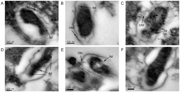
Figure 6. Detection of surface-exposed proteins using immuno-electron microscopic assay.
Vero cells infected with R. rickettsii were incubated with anti-rAdr1 (A), -rAdr2 (B), -rOmpW (C), -rPorin_4 (D), -rTolC (E), or -PBS (F) serum and subsequently immunolabeled with colloidal gold particles (10 nm) using standard procedures. The cells were then observed by transmission electron microscope. The black arrows indicate target SEPs in the inner membrane (IM) and outer membrane (OM) of R. rickettsii. Bar = 100 nm.

