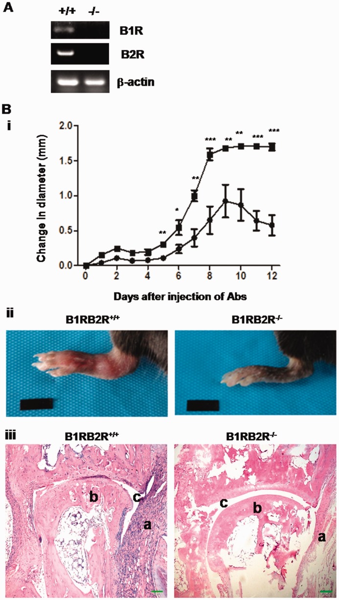
Fig. 1.
B1RB2R deficiency attenuates CAIA in mice
(A) B1R and B2R mRNA are absent in B1RB2R−/− mice. Total RNA isolated from B1RB2R+/+ (+/+) and B1RB2R−/− (−/−) mice was reversely transcribed into cDNA and subsequently analysed by PCR. The PCR products of B1R and B2R were identified by agarose gel electrophoresis. Expression of β-actin mRNA serves as the control. (B) To induce CAIA, B1RB2R+/+ and B1RB2R−/– mice received i.p. injections of anti-collagen antibodies on day 0, followed by i.p. injection of LPS on day 3 (n = 6). (i) The severity of arthritis was assessed by triplicate measurement of hind paw thickness with digital callipers (Ultra-Call Mark III, F.V. Fowler, Newton, MA, USA) every day. The change in joint diameter in millimetres from the baseline on day 0 was recorded and indicated as mean (s.e.m.). Closed box: B1RB2R+/+ mice; closed circle: B1RB2R–/– mice. *P < 0.01, **P < 0.005, ***P < 0.001. (ii) On day 12 the hind paw was photographed. (iii) On day 12 the mice were euthanized and the hind ankle joints were removed. After the joints were fixed and decalcified, they were embedded in paraffin and the paraffin sections were stained with haematoxylin and eosin. The sections were viewed and photographed under a microscope. A representative stained section shows the histopathological features of arthritis, including synovial hyperplasia, bone or cartilage erosions and mononuclear cell infiltration (original magnification 100×). a: inflamed synovial tissue; b: bone; c: joint space. Scale bar represents 50 µm. i.p.: intraperitoneal; CAIA: anti-collagen antibody-induced arthritis; LPS: lipopolysaccharide.

