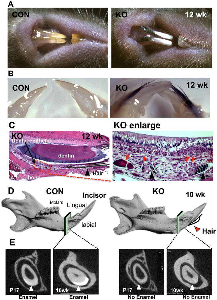
Figure 1. Deletion of Med1 generated hairs on the labial side of incisors while disrupting enamel formation.
(A, B) Hairs were generated from the labial side of chalky-colored incisors lacking enamel in Med1 KO mice (12 wk) (A) and in dissected jaws (12 wk) (B). (C) Hairs (black triangle) were observed in the tissue between the bone and the dental epithelial layer (left panel) in Med1 KO. Hairs originated from abnormal tissues (red triangles) underlying the non-polarized ameloblasts and the papillary layer (enlarged image in right panel) (12 wk). (D, E) Micro CT analysis shows the enamel hypoplasia in the Med1 KO. The 3D-reconstructed µCT images showed the structure of the mandible in CON and KO mice (10 wk) and the location of hairs (red triangle) (D). Enlarged sections of the µCT images (squares) are shown (E). The high density mineralized layer (enamel) was present on the labial side of incisors in CON incisors but absent in Med1 KO (triangles) (10 wk) with changes starting at P17. Hair was observed consistently in over 20 litters of Med1 KO mice. The entire mandibles of two mice (CON and KO) from 2 litters (10 and 12 wk) were scanned using µCT, and reproducibility was confirmed. A mineralized layer was never detected in Med1 KO incisors.

