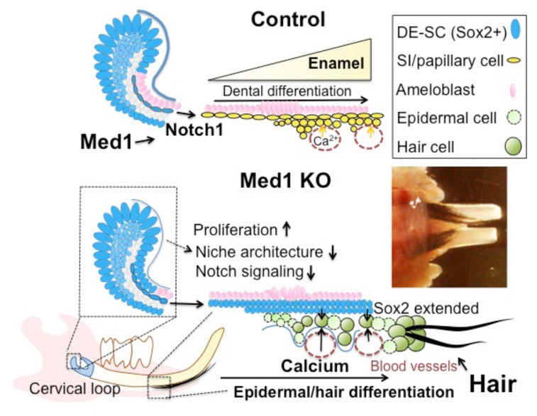
Figure 8. A proposed model in which Med1 ablation alters epithelial cell fate.
Med1 regulates Notch signaling by activating Notch1 target genes (upper diagram). Med1 maintains the niche architecture containing Sox2-expressing dental epithelial stem cells (DE-SC) (blue). Enamel is formed as dental epithelia differentiate in control incisors partly due to Notch signaling. In contrast, DE-SCs fail to commit to the dental lineage when Med1 is ablated (lower diagram). Instead, Sox2-expressing cells (blue) extend into the differentiating zones. Med1 deficient cells are exposed to extracellular calcium through the blood vessels (red-dotted circles) adjacent to the papillary layers (yellow). Med1 deficient cells are differentiated into epidermal cells (light green) and hair keratinocyte-like cells (green) and produce mature hair shafts (black) in the Med1 KO incisors (inserted picture).

