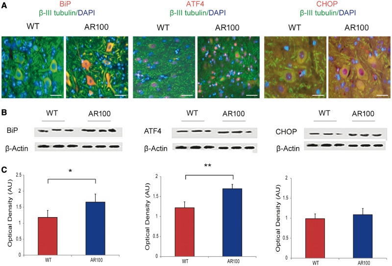
Figure 5.
Endoplasmic reticulum stress markers are elevated in the spinal cord of SBMA mice before the onset of symptoms. (A) Spinal cord lumbar 10 µM sections from AR100 and wild-type (WT) littermates at 3 months of age were then immunostained for either BiP, ATF4 or CHOP (all red) and co-stained for β-III tubulin (green), a neuronal marker and DAPI (blue), a nuclear marker. An increase in BiP and ATF4 immunoreactivity was observed in the cytoplasm of AR100 motor neurons. A modest increase in CHOP immunoreactivity was also detected. Scale bars in A = 40 µm. (B) Spinal cords from at least three different wild-type and AR100 mice at 3 months of age were analysed by western blot to quantify the levels of BiP, ATF4 and CHOP proteins. (C) Quantification by densitometry shows expression of BiP was significantly higher level in AR100 spinal cord compared to wild-type controls (n = 3, P = 0.05, t-test, n = number of mice). ATF4, was also increased in AR100 spinal cord compared to wild-type controls (n = 3, P = 0.002, t-test) but there was no significant difference in expression at 3 months of age of CHOP. Error bars show the SEM. *P < 0.05, **P < 0.01. AU = arbitrary units.

