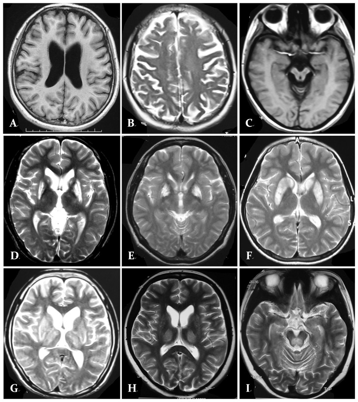
Figure 2. MRI with classically described changes in patients with Wilson.
's disease. A. Axial T1-W imaging a 18-year-old male patient shows diffuse subcortical atrophy. B. Axial T2-W imaging a 22-year-old male patient shows diffuse cortical atrophy. C. Axial T1-W imaging a 22-year-old male patient shows brainstem atrophy. D. Axial T2-W imaging a 24-year-old female patient shows bilateral putamen hyperintensity signal abnormalities. E. Axial T2-W imaging a 25-year-old male patient shows bilateral globus pallidus hyperintensity signal abnormalities. F. Axial T2-W imaging a 23-year-old male patient shows bilateral putamen and caudate nucleus hyperintensity signal abnormalities. G. Axial T2-W imaging a 20-year-old male patient shows bilateral basal ganglionic and thalamic hyperintensity in addition to diffuse atrophy . H. Axial T2-W imaging a 26-year-old female patient shows bilateral thalamic hyperintensity signal abnormalities . I. Axial T2-W imaging a 21-year-old male patient shows pons hyperintensity signal abnormalities.

