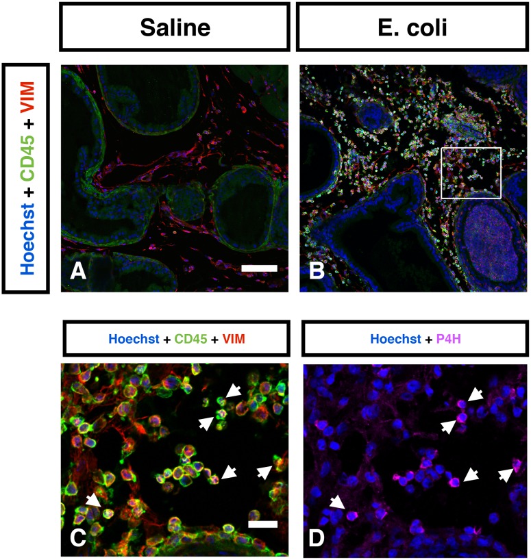
Figure 6. Identification of collagen-producing cells in the inflamed prostate.
Representative IHC images of CD45 (Green) and vimentin (Red) in the saline instilled (A) and E. coli infected (B) prostates 7 days post-instillation. n = 4–6 per group. Hoechst nuclear staining is shown in blue. Scale bar 100 µm in panel A. C. High magnification inset (box in panel B) shows abundant number of CD45+VIM+ fibrocytes in the E. coli infected prostate. Scale bar 20 µm in panel C. D. Immunostaining for prolyl 4-hydroxylase (Magneta) in the same field as panel C. White arrows indicate localization of prolyl 4-hydroxylase to CD45+VIM+ fibrocytes. Vimentin (VIM); prolyl 4-hydroxylase (P4H).

