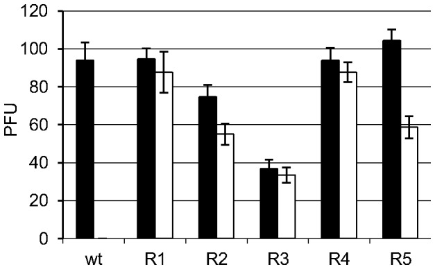
Figure 1. Antiviral activity of TSC against BVDV R1–5.

BVDV-TSCr T1–5 were propagated in MDBK cells in the absence of TSC during 20 passages. The antiviral activity of 80 µM of TSC against the viral populations obtained (BVDV R1–R5) and wt BVDV p0 was evaluated by plaque reduction assays. White and black bars indicate the number of viral plaque forming units (PFUs) formed in the presence or in the absence of TSC, respectively. (TSC vs untreated: R1 p = 0.560; R2 p = 0.054; R3 p = 0.449; R4 p = 0.382; R5 p = 0.002; p0 p = 0.003).
