
Figure 4. Multiple-step growth curve.
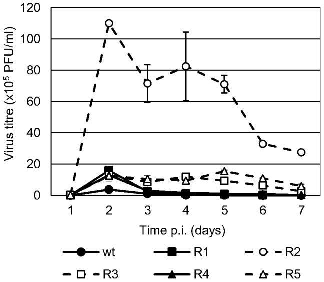
MDBK cells were infected with wt BVDV p0 or BVDV R1–5 (MOI 0.01). The viral progeny in the culture supernatant was collected every 24 h for 7 days and titrated by PFU. Viruses with altered CPE (BVDV R2, R3, and R5) are displayed in dashed lines.