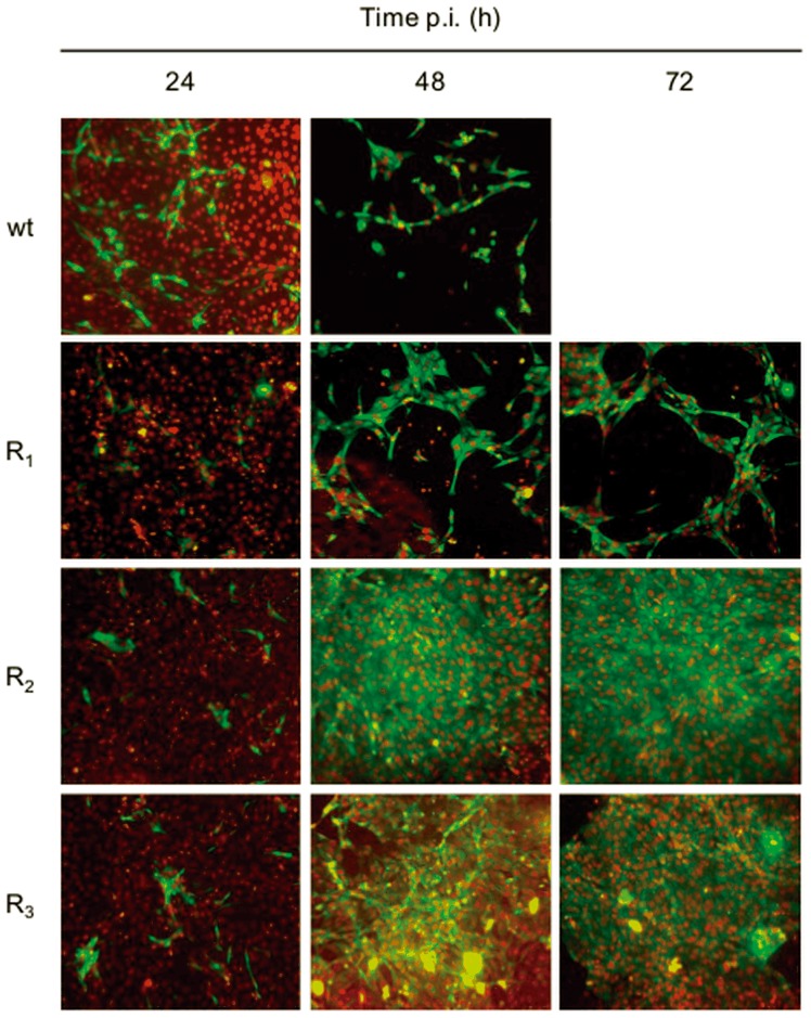
Figure 5. MDBK cells infected with wt BVDV and BVDV R viruses.
MDBK cells were infected with wt BVDV p0, BVDV R1, R2, or R3 (MOI: 0.1) and IF-stained using a monoclonal antibody against the NS3 protein at 24, 48 and 72 h p.i. The nucleus was stained with DAPI. Images were taken with a 20× objective (numerical aperture 0.40) and processed using ImageJ software (the nuclei were pseudocolored with red for better illustration).

