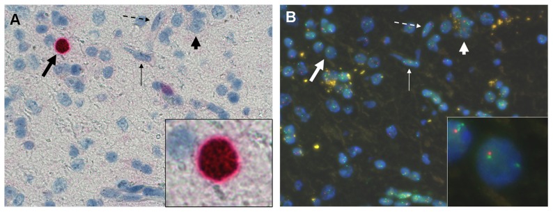
Figure 2. ImFISH interpretation.

ImFISH technique allows a simultaneous analysis of nuclear staining with MIB-1 antibody by conventional immunohistochemistry (A) and in situ hybridization with chromosomal 1p and 19q probes (B) on two separate screens. The light haematoxylin counterstaining of the immunohistochemistry step allows an easy identification of the majority of the cells analyzed: oligodendrocytes (thick arrows), astrocytes (thin arrows), neurons (short arrows) and endothelial cells (dotted arrows). Only MIB-1 labeled nuclei with an oligodendroglial morphology are analysed by FISH (framed nuclei on A and B).
