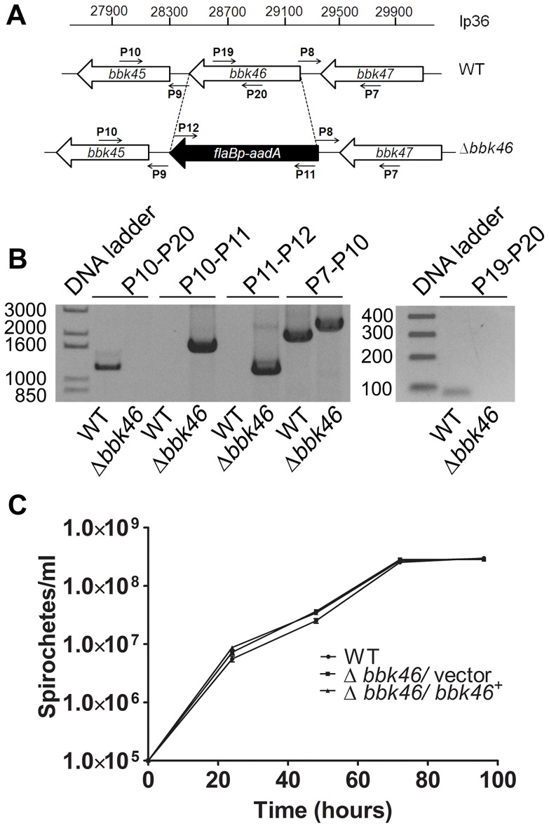
Figure 5. Generation of the Δbbk46 mutant and genetic complemented clones in B. burgdorferi.
(A) Schematic representation of the wild-type (WT) and Δbbk46 loci on lp36. The sequence of the entire bbk46 open reading frame was replaced with a flaB p-aadA antibiotic resistance cassette [37,82]. Locations of primers for analysis of the mutant clones are indicated with small arrows and labels P7-P12, P19 and P20. Primer sequences are listed in Table 5. (B) PCR analysis of the Δbbk46 mutant clone. Genomic DNA isolated from WT and Δbbk46/ vector spirochetes served as the template DNA for PCR analyses. DNA templates are indicated across the bottom of the gel image. The primer pairs used to amplify specific DNA sequences are indicated at the top of the gel image and correspond to target sequences as shown in A. Migration of the DNA ladder in base pairs is shown to the left of each image. (C) In vitro growth analysis of mutant clones. A3-68ΔBBE02 (WT), bbk46::flaB p-aadA/ pBSV2G (Δbbk46/ vector) and bbk46::flaB p-aadA/ pBSV2G-bbk46 (Δbbk46/ bbk46+) spirochetes were inoculated in triplicate at a density of 1×10⁵ spirochetes/ml in 5 ml of BSKII medium. Spirochete densities were determined every 24 hours under dark field microscopy using a Petroff-Hausser chamber over the course of 96 hours. The data are represented as the number of spirochetes per ml over time (hours) and is expressed as the average of 3 biological replicates. Error bars indicate the standard deviation from the mean.

