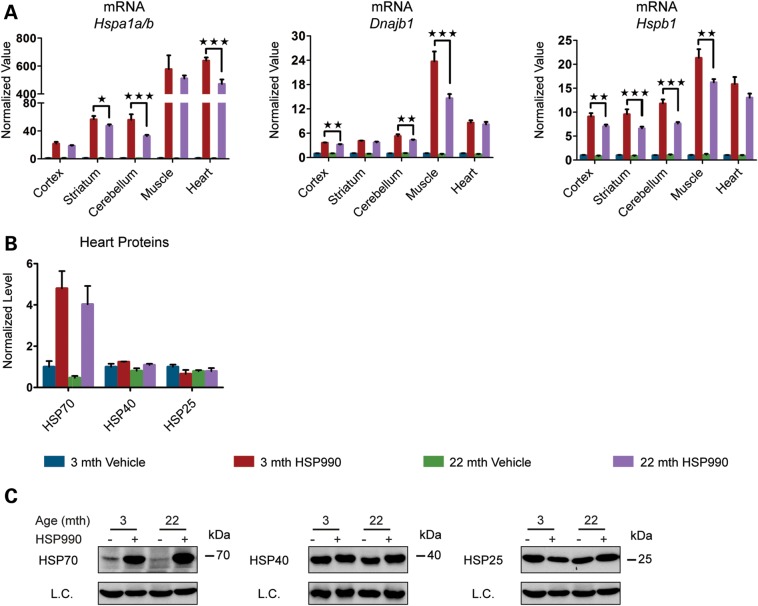
Figure 5.
Analysis of the dynamics of HSP induction. Tissues were taken from 3- and 22-month-old mice 4 h after treatment with HSP990 (12 mg/kg) or vehicle. (A) qPCR analysis of the expression levels of Hspa1a/b, Dnajb1 and Hspb1 in brain regions and peripheral tissues. Values were calculated relative to vehicle-treated young mice. (B) Relative protein level of HSP70, HSP40 and HSP25 in the heart. Densitometric values were calculated relative to vehicle-treated young mice. (C) Representative western blots for HSP70, HSP40 and HSP25. Data are the mean ± SEM. 3 < n < 6/group. *P < 0.05; **P < 0.01; ***P < 0.001. Asterisk indicates the statistically significant difference in the level of induction. L.C., loading control; mth, months.

