Figure 3.
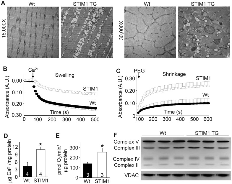
STIM1 overexpression leads to a mitochondrial pathology in muscle. (A) Electron microscopic images of Wt and STIM1 TG quadriceps muscle at two different magnifications. (B) Light scattering assessed in mitochondria suspension during a Ca2+ stimulus to induce swelling derived from Wt (solid) and STIM1 TG (clear) muscle. (C) Same assay as in “B” except that mitochondria shrinking was assessed in response to PEG addition. (D) Quantitation of mitochondrial matrix Ca2+ content in isolated mitochondria from Wt and STIM1 TG muscle. (E) Analysis of baseline O2 consumption in mitochondria isolated from Wt and STIM1 TG muscle. (F) Western blot analysis for the indicated mitochondrial proteins in Wt and STIM1 TG muscle protein lysates. *P < 0.05 compared with Wt mice and the number of animals used is shown in each panel in the bars.

