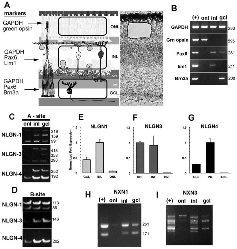
Figure 4.

Laminar position of alternatively spliced neuroligin and neurexin transcripts in the chick retina. A: The retina is compartmentalized into outer and inner nuclear layers and the ganglion cell layer, each separated by the synaptic plexiform layers. A representative laser capture microdissection cut of the outer nuclear layer is indicated here. B: LCM samples were validated for purity and lack of contamination of RNA from other retinal layers by PCR using primers specific for photoreceptors (green opsin), inner nuclear layer horizontal cells (Lim1), inner retina (Pax6), or the ganglion cells (Brn3). Detection of NLGN1, -3, or -4 alternatively spliced variants in isolated nuclear layers is revealed by analysis with primers flanking splice sites -A (C) or -B (D). E–G: Real-time PCR was used to show relative abundance of NLGN1, -3 and -4 transcripts in relation to the housekeeping gene GAPDH. Different sized alternative spliced NRXN1 (H) and NRXN3 (I) transcripts reveal differences in splicing.
