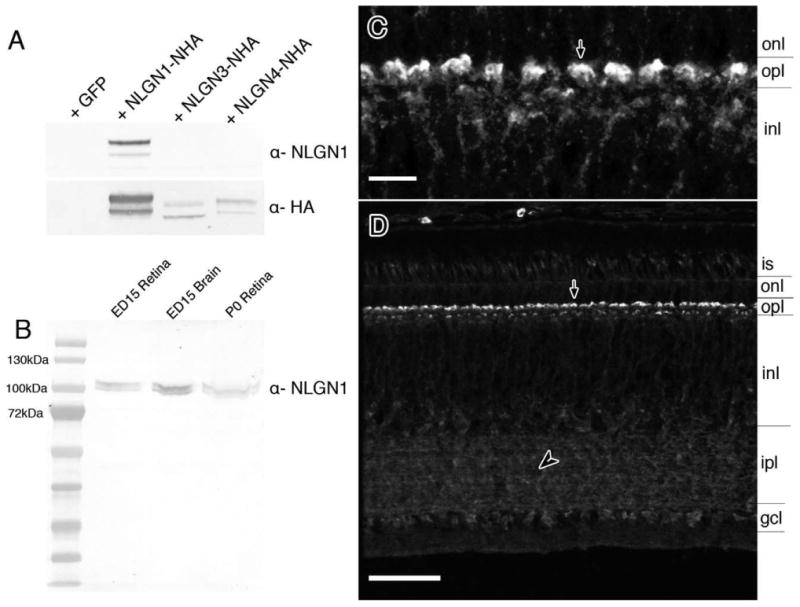
Figure 5.

Detection of NLGN1 protein in the chick retina and antibody specificity. A: Western blots with HA-tagged recombinant chicken NLGN1, -3, or -4 proteins were probed with NLGN1 antibodies to demonstrate a lack of cross-reactivity to other NLGN family members. Samples run in parallel were probed with anti-rat HA antibodies as a loading control. B: Native protein from ED15 and P0 chick retinas and ED15 chick forebrain were separated by SDS-PAGE gel electrophoresis and probed with antibodies against NLGN1. C,D: Tissue sections from ED20 retinas were probed with NLGN1 antibodies and detected by fluorescence immunohistochemistry. Panel C is a magnified image of the outer plexiform layer with NLGN1 positive dendrites projecting from the outer inner nuclear layer into the outer plexiform layer (arrow). is, photoreceptor inner segment; onl, outer nuclear layer; opl, outer plexiform; inl, inner nuclear layer; ipl, inner plexiform layer; gcl, ganglion cell layer. Scale bars = 5 μm in C; 25 μm in D.
