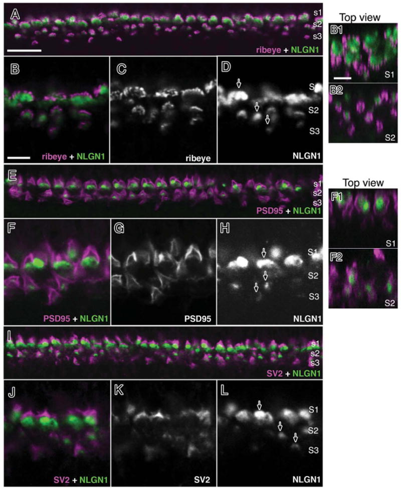
Figure 7.

NLGN1 expression adjacent to photoreceptor terminals in the mature chick retina. Double-labeled immunohistochemistry for NLGN1 and ribeye (A–D), PSD95 (E–H) or SV2 (I–L) is shown in a 21-day-old (ED21) chick retina at the level of the outer plexiform layer. Accumulation of NLGN1 and presynaptic proteins in three distinct strata (S1–3) showed separate labeling patterns between NLGN1 immunoreactive bodies and the boundaries of photoreceptor terminals. Single arrows indicate the ribbed appearance of synaptic ribbon-like structures (D), the peripheral staining of PSD95 (E), or the SV2 positive vesicle pool (I) of double cone terminals. Panels to the right (B1,B2,F1,F2) are top-down orthogonal views showing size differences between synaptic terminals in S1 and S2. Scale bars = 15 μm in A; 5 μm in B,B1.
