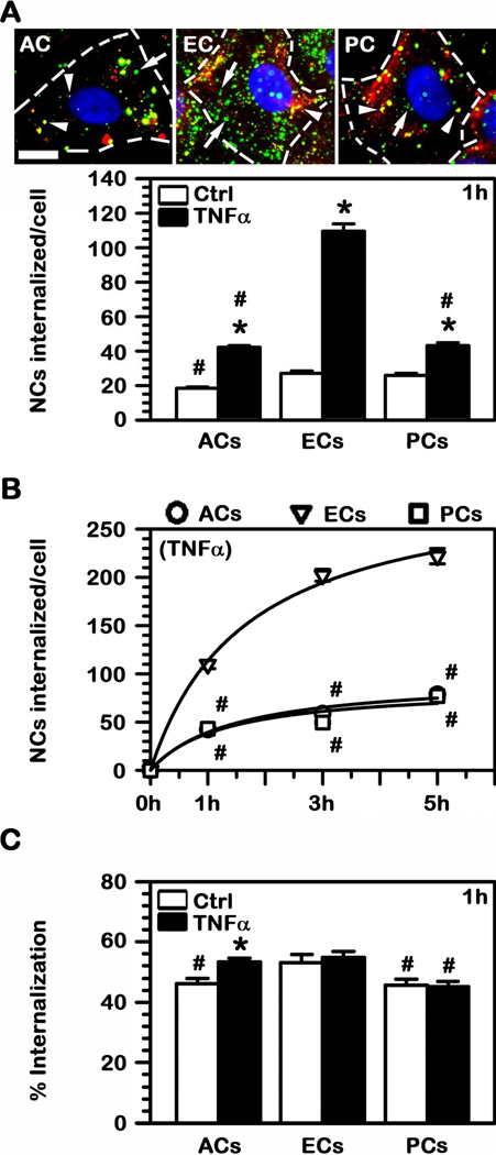
Fig. 3.
Internalization of anti-ICAM NCs into BBB cells. (A) FITC-labeled anti-ICAM NCs were incubated with TNFα-activated (images) or control human brain astrocytes (ACs), endothelial cells (ECs), or pericytes (PCs) for 1 h at 37°C. Non-bound carriers were washed, cells were fixed, and surface-located particles were immunostained with a Texas Red-secondary antibody (green FITC + Texas Red = yellow particles; arrowheads) versus internalized counterparts which remain single-labeled in green FITC (arrows). Scale bar = 10 μm. The absolute number of nanocarriers internalized per cell after 1 h incubation or (B) during a period of 5 h were quantified by fluorescence microscopy. (C) The percent of cell-associated particles that were internalized by cells was quantified as in (A). (A–C) #Comparison to ECs within each condition; *comparison between TNFα and control for each cell type (p<0.05 by Student’s t-test).

