Figure 3.
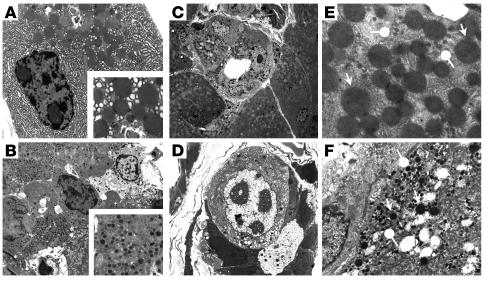
Ultrastructural analysis of E2F1/E2F2 double-homozygote pancreas sections. Exocrine (A) and endocrine (B) cells in pancreata from WT mice. Insets show the aspect of normal acinar and β cell granules. Pancreas from a 3-month-old male DKO mouse (C_F). Ductal structures composed of transitional cells with ductal cell features, but containing zymogen granules (C), or both zymogen and endocrine granules, can be observed in DKO mice (D). Cells containing both types of granules (pointed arrows, acinar granules; round arrows, endocrine granules) (E and F). Original magnification: (A, B, E, F) ∞3,400; (C) ∞1,100; (D) ∞2600; insets in A and B, ∞10,500.

