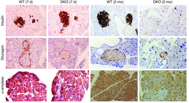
Figure 4.
Immunohistochemical analysis of expression of pancreas-specific protein markers. Shown are representative pancreas sections of 7-day-old and 2-month-old WT and DKO male mice immunostained with Ab’s to insulin, glucagon, and α-amylase. A light hematoxylin counterstaining was performed in all sections (insulin and glucagon, ∞400; α-amylase, ∞200). Similar results were obtained when DKO female mice were analyzed (data not shown).

