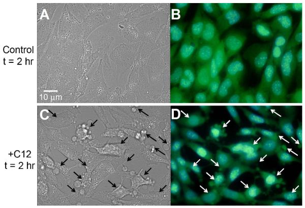
Fig. 2. C12 causes cell and nuclear shrinkage in WT MEF.
WT MEF were observed under DIC (A, C) or fluorescence (B, D) optics at 2 hr. Fluorescence images of blue Hoechst-stained nuclei and green BCECF-stained cytosol were overlain using Adobe Photoshop. Control, untreated cells showed typical, flattened morphology with oval nuclei and BCECF distributed throughout the cytosol and nuclei. Compared to control conditions, C12-treated WT cells and nuclei were obviously shrunken, and plasma membrane had multiple blebs (arrows) containing BCECF. Images typical of >15 each in three different coverglasses each.

