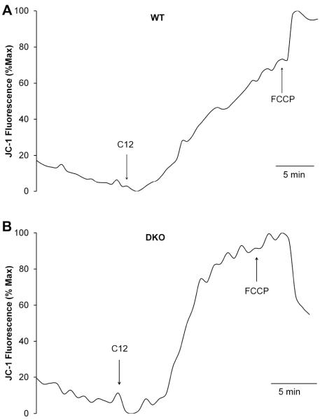
Fig. 7. Effects of C12 on Δφmito in WT and DKO MEF.
MEF were loaded with JC1 then mounted on the imaging microscope for measurement of fluorescence under control conditions and during treatment with C12 (50 μM) and FCCP (10 μM). JC1 fluorescence showed steady value in control conditions and slow increase (equivalent to depolarization) in response to C12 in both WT (A) and DKO (B) MEF. FCCP caused maximal depolarization of Δφmito at the end of the experiments. Results typical of 3-5 experiments each.

