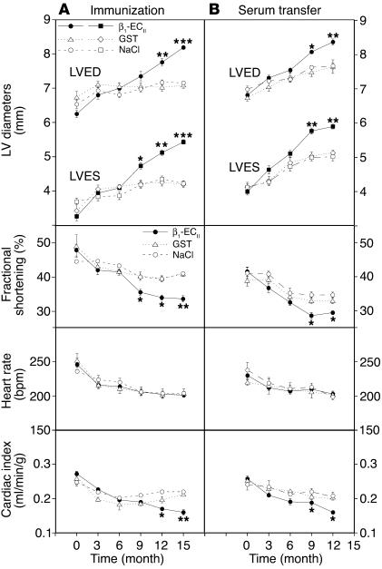
Figure 5.
Echocardiographic follow-up. (A) Immunization and (B) transfer experiment. The panels depict the time course of selected echocardiographic parameters. Upper panels: LVED and end-systolic diameters (LVES). Middle panels: fractional shortening (%), derived from (LVED-LVES/LVED ∞ 100), and heart rate (bpm). Lower panels: Cardiac index corresponding to CO/BW. CO (milliliters per minute) was assessed by echocardiography (see Methods). Error bars indicate mean plus or minus SEM.*P < 0.05; **P < 0.01; ***P < 0.001 (ANOVA and Bonferroni post hoc test).

